They have created a radiotherapy treatment room on a computer. Radiotherapy is used to treat people with cancer. While the patient lies on a couch, pencil thin beams of radiation are pointed at them. The beams are lined up very carefully so they intersect inside the patient at the site of the tumour. The high level of radiation kills off the cancer cells. But its a precise art. If the beams are not pointed precisely at the tumour the cancerous cells will be left intact and healthy tissue will be destroyed instead. Vital organs like the spine, the kidneys or the liver could be damaged. That is why its so important that health professionals who administer radiotherapy are good at what they do. Unfortunately, until now, there have been few opportunities for them to hone their skills. There are no spare radiotherapy treatment suites in the UK where they can practise. 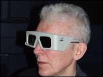 | | The 3D glasses |
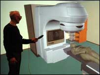
But experts at Hull University and The Princess Royal Hospital have solved the problem. They have written computer software that simulates a radiotherapy treatment room. A lifesize picture of the computer generated room is projected onto a wall in a classroom. The picture shows a couch, with a patient lying on it. Inside the patient you can see the main organs, which are colour coded. You can also see the tumour. The picture shows cupboards on the walls of the treatment room and it shows the moveable machinery that generates the beams of radiation.
The students put on 3D glasses and the whole image springs to life. The students can walk to and fro in front of the image and move around the patient in three dimensions, seeing them from different angles. They can also control the radiotherapy equipment in the picture. Later, the computer shows them the results of their work. Did they get the treatment right? Bodyscan images from real patients are used throughout. The virtual reality radiotherapy treatment suite has been developed at the Hull Immersive Visualisation Environment at Hull University. Professor Roger Phillips, the leader of the project, told us " The University of Hull is in the process of developing a consortium with other universities and manufacturers to investigate how our equipment can be used to the full. Aarhus University Hospital in Denmark, Hertfordshire University Hospital and the University of Central England are currently replicating this technology and building their own training suites ". This new radiotherapy training facility will be used initially by medical students at Hull University and by staff at the Princess Royal Hospital. . |